Medical OSCES

Medical OSCES for medical students and PG Doctors

Menu
  • Home
  • Static Page
  • Radiology Cases
    • Neurology
    • Peadiactric
    • Chest
    • GIT
    • GUT
  • Radiology Notes
    • Neurology
    • Peadiactric
    • Chest
    • GIT
    • GUT
  • Lectures
    • Radiology
    • Anatomy
    • Physics
    • Radiography
  • Videos
  • Books
  • Surprise Me
Pediatric EMCQs Pediatric EMCQ 02 : Skin lesions

Pediatric EMCQ 02 : Skin lesions

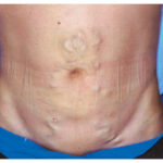
image
A. Café au lait spots
B. Capillary haemangioma
C. Port-wine stain 
D. Shagreen patch
E. Adenoma sebaceum
F. Cutis Marmorata
G. Herald's patch
H. Peri-oral pigmentation
I. Hypo pigmentation
J. Tuber


Select one option from the list above that is most suitable for the following patients

1) An 11 year old boy with learning difficulties has an acne type rash over the nose

2) A 4 year old boy with a history of focal seizures and a birth mark

3) An infant with Down's syndrome has mottled appearance to the skin.


Answers …E C F

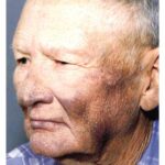
E. Adenoma sebaceum
image

Note:
relates to the condition known as Tuberous sclerosis. It is a condition where by hamartomatous lesions become evident in early childhood. Seizures and learning difficulties are common and skin manifestations include fibrous angiomatous lesions in the naso labial folds known as adenoma sebaceum. White macules are seen over the trunk known as Ash leaf macules and Shagreen patches resemble goose like flesh. Subungual fibromata are also common. 

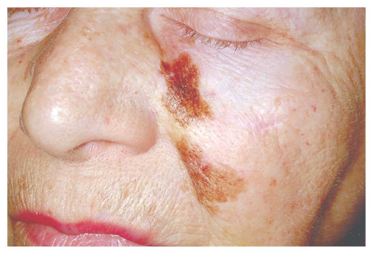
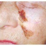
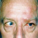
C. Port-wine stain 
image
Note:
relates to Sturge-Weber syndrome. The association and localization of aberrant vasculature in the facial skin, eyes and meninges. Most commonly in the trigeminal nerve distribution.

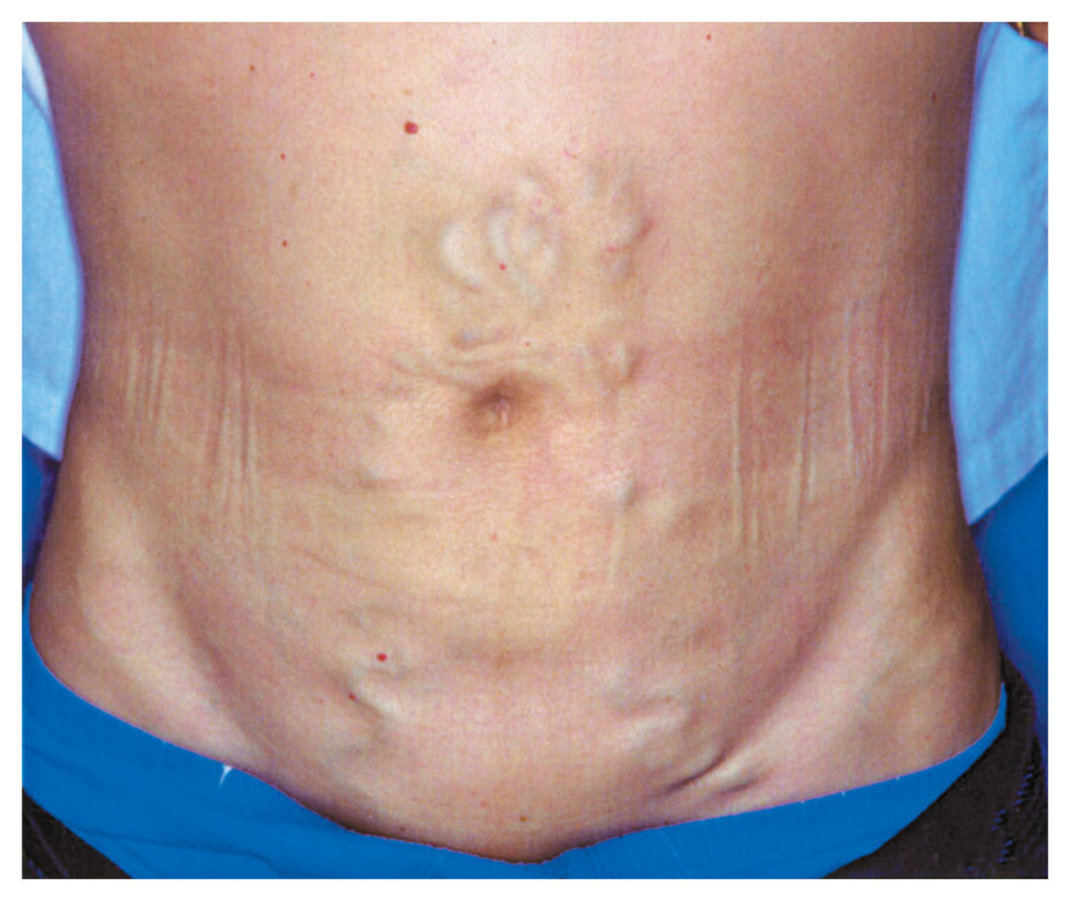
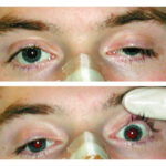
F. Cutis Marmorata
Note: image
relates to Cutis Marmorata, a mottled, marbled type appearance to the skin. Common in Down's syndrome as well as other Trisomies, hypothyroidism and Cornelia-de-Lange syndrome.
Add Comment
Pediatric EMCQs
  • Tweet
  • Share
  • Share
  • Share
  • Share

Related Posts

Post a Comment

Total Tayangan Laman

  • …

  • …

Weekly Posts

  • Diffuse Axonal Injury
    Diffuse Axonal Injury
    Findings: Diffuse brain swelling Focal punctate hemorrhage scattered in the white matter, corpus callosum, and brain stem Right subgaleal...
  • Adrenal adenoma
    Adrenal adenoma
    •Findings: –Large right adrenal lesion –Isointense to liver on in-phase scan –Hypointense to liver (signal loss) on out-of-phase scan ...
  • SVC obstruction
    SVC obstruction
    •Findings: –Contrast-enhanced CT shows intense enhancement of the medial segment of the left lobe = “liver hot spot” sign –Collateral ve...
  • Name the arrowed structure ?
    Name the arrowed structure ?
    Left Ventricle

Label

  • Cardiology (12)
  • Chest x-ray (9)
  • CT Scans (6)
  • Dermatology (56)
  • ECG (2)
  • Endocrinology (10)
  • ENT (2)
  • Gastroenterology (15)
  • Genetics (6)
  • Gyn and Obs (5)
  • gynaecology (4)
  • Haematology (17)
  • Image Challenge (214)
  • Immunology (3)
  • infectious and parasitic disease (3)
  • Infectious disease (5)
  • Instruments (1)
  • Microbiology (2)
  • Musculoskeletal (3)
  • NEJ Cases (9)
  • Nephrology (2)
  • Neurology (15)
  • oncology (3)
  • Ophthalmology (13)
  • Orthopaedic (8)
  • Parasitology (4)
  • Peadiactrics (12)
  • Pediatric EMCQ (1)
  • Pediatric EMCQs (5)
  • Pharmacology (4)
  • Pulmonology (9)
  • Radiology (30)
  • Real Cases (5)
  • Rheumatology (7)
  • Skull X-ray (2)
  • Surgery (4)
  • Vascular surgery (1)
  • x-ray (6)

Contact

Name

Email *

Message *

Copyright © 2014 Medical OSCES All Right Reserved
Blogger Templates Created by Arlina Design